- Intraoral scanning records the shape of your teeth and gums with a small camera, without impression material or trays.
- The scanner uses light, not radiation, and the procedure is painless.
- A full arch usually takes a few minutes, and the result appears immediately on screen as a 3D model.
- For crowns, veneers, inlays, onlays and work on a few teeth, studies show accuracy comparable to a good-quality conventional impression.
- Scanning has limits too: subgingival preparations and areas covered by blood or saliva must be prepared first, because the scanner only records what it can see.
What is intraoral scanning?
For almost any prosthetic work, the dental technician needs an exact copy of the patient's teeth. Traditionally this copy is taken with an impression: a tray filled with impression material is held in the mouth for a few minutes until the material sets.
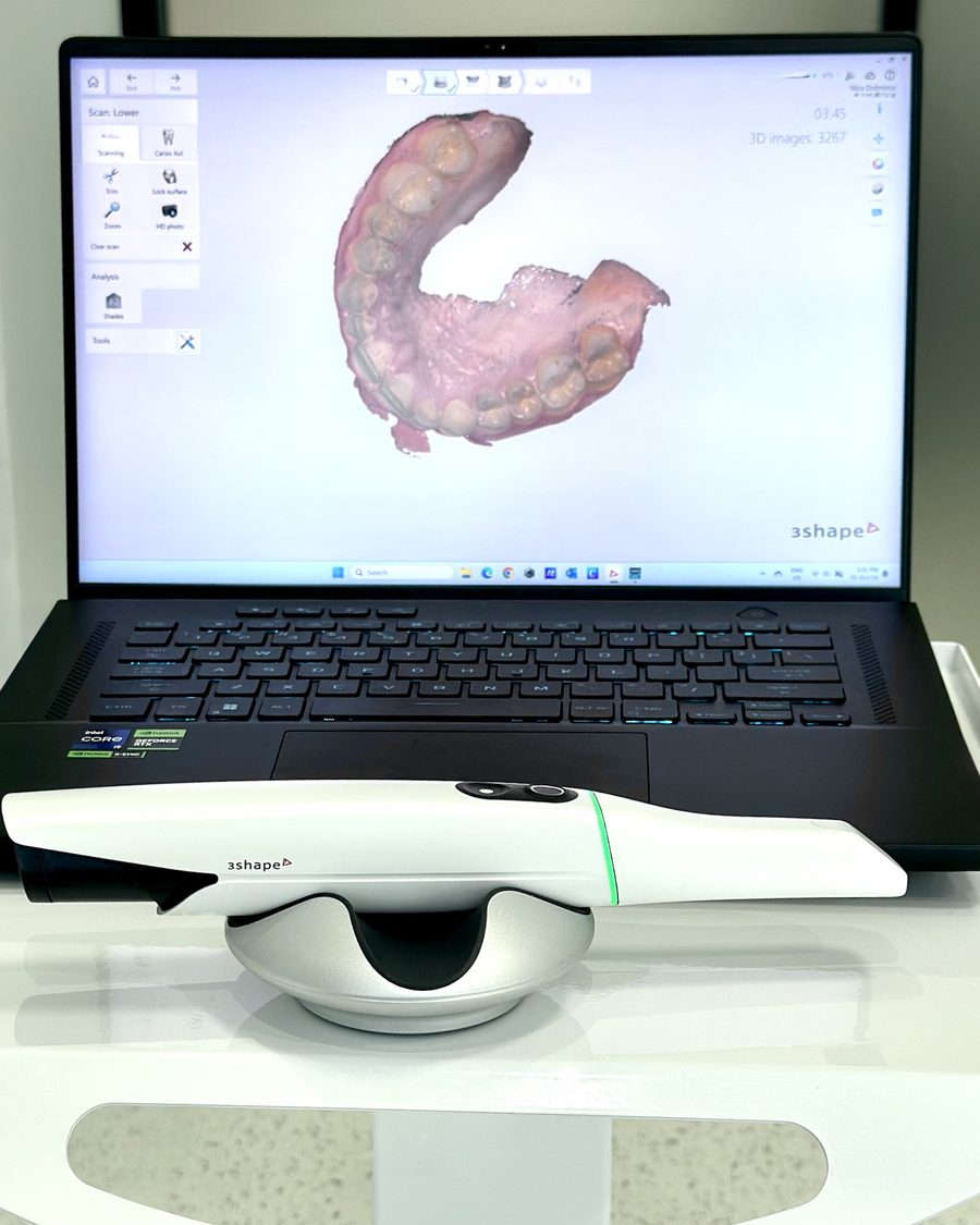
Intraoral scanning captures the same information digitally. A scanner roughly the size of an electric toothbrush is moved along the teeth and records thousands of images per second. The software assembles them into a 3D model of the teeth and gums, which appears immediately on screen. This file is called a digital impression.
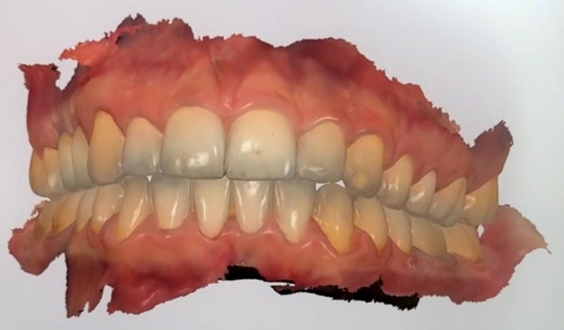
How is the scan done?
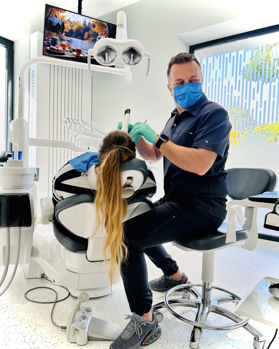
- Preparation. The teeth are gently dried with air so that saliva does not interfere. If the preparation is subgingival, the gum is carefully moved aside so that the preparation margin is visible.
- Scanning. I move the scanner along the arch, over the cheek side, the tongue side and the biting surfaces. You can breathe and swallow normally, and pauses are not a problem.
- The bite. With your teeth together, I scan the side of the teeth to record how the arches meet.
- Checking on screen. I examine the magnified 3D model. If an area is not clear enough, I rescan it on the spot, without repeating the whole procedure.
- Sending to the laboratory. The file reaches the dental technician digitally, with no physical impression to transport.
What are the benefits over conventional impressions?
- Comfort. No tray of impression material held in the mouth, so no taste of the material and no gagging, which for many patients is the most unpleasant part of treatment.
- Immediate control. I see every detail of the preparation magnified on screen. A problem is corrected on the spot, not after the work has come back from the laboratory.
- No distortion over time. Impression materials can shrink or distort if they are not cast in time. The digital file stays exactly the same.
- Better communication with the laboratory. The technician receives the model immediately and can ask questions the same day.
- A record. Scans are stored, so they can be compared over time, for example to monitor tooth wear or gum recession.
- Clearer explanations. I can show you on screen, in 3D, exactly what is wrong with a tooth and what I suggest we do.
How accurate is a digital impression?
For work on one tooth or a few teeth, such as crowns, veneers, inlays, onlays and short bridges, clinical and laboratory studies show accuracy comparable to good-quality conventional impressions. The marginal fit of restorations made from scans is within clinically accepted limits.
For full arches, especially extensive work on several implants, accuracy depends greatly on technique, scanner and the clinical situation. In some of these cases the dentist may combine scanning with other verification methods. The choice is made case by case.
Accuracy does not depend on the device alone. Correct tooth preparation, control of saliva and bleeding, and the experience of the person scanning matter just as much.
What are the limitations of scanning?
The scanner only records what it can see. That is why:
- subgingival preparations must be exposed first, usually with a retraction cord, exactly as for a conventional impression
- blood and saliva hide detail and must be controlled
- in patients with limited mouth opening, access to the molar area can be more difficult
None of these limitations is unique to scanning: a conventional impression needs the same conditions to be accurate.
Which scanner do I use?
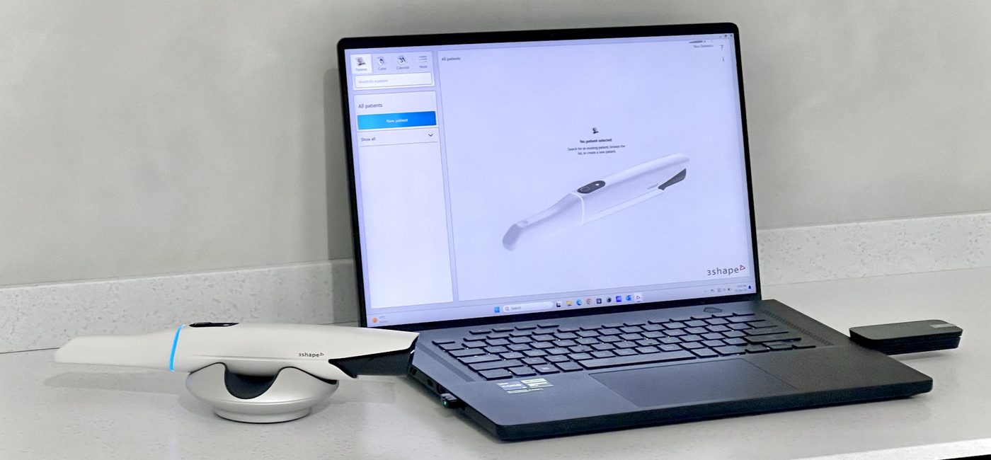
At ARTeeth Dental Clinic I work with the 3Shape TRIOS 5 intraoral scanner. The tips that go into the mouth are sterilised after every patient.
You can read more about the 3Shape TRIOS 5 intraoral scanner at ARTeeth Dental Clinic on the clinic's website (in Romanian).
Where is intraoral scanning used?
- crowns, veneers, inlays and onlays
- bridges and implant restorations
- digital smile design and aesthetic mock-ups
- night guards for bruxism
- orthodontic treatment with clear aligners
- recording the starting situation before complex treatment
Frequently asked questions
Does intraoral scanning hurt?
No. The scanner simply glides along the teeth without pressing on them. Most patients find it far more comfortable than a conventional impression.
Is a scan the same as an X-ray?
No. An intraoral scanner uses light to record the surface of the teeth and gums. It emits no radiation and does not replace X-rays, which show the inside of the tooth and the bone.
How long does a scan take?
A full arch usually takes a few minutes. The exact time depends on how many teeth need to be recorded and on the planned treatment.
Is it as accurate as a conventional impression?
For crowns, veneers, inlays, onlays and work on a few teeth, studies show accuracy comparable to a good-quality conventional impression. For some extensive full-arch work, the dentist may choose to combine methods.
Can I see the result of the scan?
Yes. The 3D model appears on screen in real time, and after the scan I can show you every tooth magnified.
Is scanning hygienic?
Yes. The scanner tips that go into the mouth are sterilised after every patient.
Is a conventional impression ever still needed?
Rarely, but yes. In certain situations, such as some complex full-arch work or very limited mouth opening, a conventional impression or a combination of methods may be the better choice.
This article provides general information and does not replace a consultation and clinical examination. The right impression method is chosen after assessing each case.

Dentist with over 20 years of practice dedicated to prosthodontics and dental aesthetics. Founder of ARTeeth Dental Clinic, Bucharest.
At your first consultation I examine your teeth and, if needed, take an intraoral scan. You will see the 3D model on screen and we will discuss the treatment options together.